
Visual anatomy employs visual aids – like atlases – to understand the human body‚ treating it like a geographical map with organs as ‘countries’․
The Importance of Visual Learning in Anatomy
Visual learning is crucial in anatomy because of the body’s complexity; anatomy atlases greatly aid comprehension․ Studying the human form benefits immensely from visual aids‚ enabling a deeper understanding of structures and their relationships․
Approaching the body as a ‘map’ – continents representing body parts‚ organs as ‘countries’ – fosters intuitive spatial awareness‚ benefiting all learners‚ especially those with visual impairments․
Anatomy Atlases: A Historical Overview
Historically‚ anatomy relied on detailed illustrations‚ evolving into modern anatomy atlases․ These resources transitioned from artistic renderings to increasingly realistic depictions‚ mirroring the body’s intricate details․
Contemporary atlases‚ like 3D models‚ now present the body as an explorable ‘map’‚ dividing it into regions and organs‚ enhancing spatial understanding and offering interactive visual experiences․
Skeletal System – The Framework
Visualizing the skeletal system reveals its foundational role‚ akin to a building’s structure‚ supporting the body and defining its overall form and movement․
Visualizing Bone Structure and Types
Bone structure‚ when visualized‚ resembles architectural designs – compact bone as dense walls‚ and spongy bone as internal support beams․ Different bone types – long‚ short‚ flat‚ irregular‚ and sesamoid – can be understood through analogies; long bones are like pillars‚ flat bones like shields‚ and irregular bones like uniquely sculpted forms․
Key Anatomical Landmarks: A Regional Approach
A regional approach to landmarks utilizes the body as a map․ The head and neck are a ‘mountain range’ with specific peaks (bony prominences)․ The thorax is a ‘protected valley’ housing vital organs․ Limbs are ‘roadways’ with arteries as major routes․ Visualizing regions simplifies complex anatomy‚ fostering spatial understanding․

Muscular System – Movement and Support
Muscles are visualized as ‘engines’ powering movement‚ with attachments as ‘cables’․ Muscle groups function as coordinated ‘teams’‚ enabling complex actions throughout the body․
Muscle Groups and Their Functions (Visual Breakdown)
Visualizing muscle groups involves understanding them as specialized units․ For example‚ consider the biceps as a ‘lifting team’ and the quadriceps as a ‘kicking force’․
Each group’s function – flexion‚ extension‚ abduction‚ adduction – can be represented as directional arrows on a diagram․ Detailed anatomical charts showcase origins‚ insertions‚ and actions‚ clarifying how these ‘teams’ collaborate to produce movement․
This breakdown simplifies complex interactions‚ fostering a deeper comprehension of musculoskeletal mechanics․
Understanding Muscle Attachments and Actions
Muscle attachments – origins and insertions – can be visualized as ‘anchor points’ for movement․ Imagine tendons as strong ropes connecting muscles to bones‚ facilitating force transmission․
Actions‚ like flexion or extension‚ are best understood as directional pulls․ Diagrams illustrating these ‘pulls’ clarify how muscles create motion at joints․
Visualizing these connections demystifies complex biomechanics‚ revealing how muscles work as levers․

Nervous System – Communication Network
The nervous system resembles a complex network of electrical wires‚ transmitting signals throughout the body‚ much like a sophisticated communication system․
Brain Anatomy: A Visual Guide to Lobes and Structures
Visualizing the brain involves understanding its distinct lobes – frontal‚ parietal‚ temporal‚ and occipital – each with specialized functions․ Think of the brain as a control center‚ with each lobe managing specific ‘departments’․ Detailed 3D models and anatomical illustrations are crucial for grasping the intricate structures within‚ like the cerebellum and brainstem‚ essential for coordination and vital functions․ These visual guides aid in comprehending the brain’s complex organization and its role in controlling bodily functions and cognitive processes․
Spinal Cord and Peripheral Nerves: Mapping the Pathways
Visualizing the spinal cord and peripheral nerves requires understanding them as the body’s ‘communication network’․ The spinal cord acts as the central highway‚ transmitting signals between the brain and body․ Peripheral nerves branch out like roads‚ reaching limbs and organs․ Anatomical charts effectively illustrate these pathways‚ showing how signals travel for sensation and movement‚ crucial for understanding neurological function and potential injury locations․

Cardiovascular System – The Life Force
Visualizing the cardiovascular system reveals the heart as a central pump‚ with arteries and veins acting as a network of rivers delivering life-sustaining oxygen․
Heart Anatomy: Chambers‚ Valves‚ and Circulation
Visualizing the heart’s anatomy clarifies its function: chambers act as receiving and pumping rooms‚ while valves ensure unidirectional blood flow․ Imagine a well-managed train station – atria receive‚ ventricles dispatch․
The circulatory pathways‚ arteries carrying oxygenated blood away‚ and veins returning deoxygenated blood to the heart‚ resemble a complex railway system efficiently transporting vital resources throughout the body․ Understanding these structures is crucial․
Blood Vessels: Arteries‚ Veins‚ and Capillaries (Visual Tracing)
Visualizing blood vessels as a delivery network enhances understanding․ Arteries‚ like major highways‚ transport blood from the heart․ Veins‚ the return routes‚ bring blood to the heart․
Capillaries‚ tiny side streets‚ facilitate exchange with tissues․ Tracing this network reveals how oxygen and nutrients reach cells‚ and waste products are removed – a remarkably efficient logistical system within the body;
Respiratory System – Breath of Life
Visualizing lungs and airways reveals an inverted tree structure‚ branching to maximize surface area for efficient oxygen exchange – the ‘breath of life’․
Lungs and Airways: A Visual Exploration
Detailed visuals showcase the lungs’ branching network‚ resembling an upside-down tree‚ with the trachea as the trunk and bronchioles as smaller branches․ This structure dramatically increases surface area․
Exploring the airways – nasal cavity‚ pharynx‚ larynx‚ trachea‚ bronchi‚ and bronchioles – reveals a pathway for air․ Visual guides highlight how air travels‚ enabling efficient oxygen uptake and carbon dioxide removal‚ vital for sustaining life․ Understanding this pathway is crucial for comprehending respiratory function․
Diaphragm and Breathing Mechanics
Visualizing the diaphragm – a dome-shaped muscle – clarifies its pivotal role in respiration․ Contraction flattens the diaphragm‚ increasing chest volume and drawing air into the lungs‚ akin to pulling a syringe․
Relaxation restores the dome shape‚ decreasing volume and expelling air․ Interactive diagrams demonstrate how intercostal muscles assist‚ expanding and contracting the rib cage․ This coordinated action‚ visually represented‚ explains the mechanics of inhalation and exhalation․
Digestive System – Fueling the Body
Visualizing the gastrointestinal tract as a disassembly line‚ from mouth to anus‚ clarifies nutrient extraction; accessory organs support this process effectively․
Gastrointestinal Tract: From Mouth to Anus (Visual Journey)
Imagine the GI tract as a continuous‚ winding pathway․ Starting with the mouth – the entry point – visualize food being mechanically and chemically broken down․
Follow its journey through the esophagus‚ stomach (a churning mixer)‚ small intestine (nutrient absorption central)‚ large intestine (water reclamation)‚ and finally‚ elimination via the anus․
Visualizing each segment’s unique structure and function clarifies the entire digestive process‚ emphasizing its sequential‚ coordinated nature․
Accessory Organs: Liver‚ Pancreas‚ and Gallbladder
Picture these organs as support stations alongside the GI tract’s main highway․ The liver‚ a processing plant‚ filters blood and produces bile․
The pancreas‚ a dual-function factory‚ releases digestive enzymes and hormones․ Visualize the gallbladder as a bile storage reservoir‚ releasing it when needed for fat digestion․
Understanding their interconnected roles clarifies how they collectively aid in efficient nutrient breakdown and absorption․

Endocrine System – Chemical Messengers
Visualize endocrine glands as postal offices‚ releasing hormones (messages) into the bloodstream to regulate body functions‚ creating feedback loops․
Major Endocrine Glands and Their Hormones (Visual Representation)
Imagine the pituitary as a central command center‚ overseeing hormone release․ The thyroid‚ a factory producing metabolism regulators․ Adrenal glands‚ emergency response units releasing cortisol․
Visualize the pancreas as a sugar-balancing station‚ secreting insulin․ Ovaries/testes‚ reproductive hormone producers․ A visual map linking each gland to its specific hormonal ‘products’ clarifies their roles․
Hormonal Regulation and Feedback Loops
Picture a thermostat controlling room temperature – that’s hormonal regulation! High hormone levels signal a ‘decrease’ command‚ like cooling a warm room․ Conversely‚ low levels trigger ‘increase’‚ similar to heating a cold one․
Visualize these loops as interconnected circuits‚ maintaining balance․ Negative feedback is the thermostat; positive feedback amplifies a signal‚ like childbirth contractions․
Immune System – Body’s Defense
Envision the lymphatic system as a network of roads (vessels) with checkpoints (nodes) where immune cells patrol‚ defending against invaders․
Lymphatic System: Vessels‚ Nodes‚ and Organs (Visual Map)
Imagine the lymphatic system as a complex drainage system alongside blood vessels․ Visualize delicate vessels collecting fluid‚ flowing towards nodes – bean-shaped filters acting as security checkpoints․
Picture these nodes strategically positioned throughout the body‚ teeming with immune cells․ See organs like the spleen and thymus as central command centers‚ orchestrating immune responses․ This ‘visual map’ highlights how fluid travels‚ gets filtered‚ and immunity is maintained․
Immune Cells and Their Functions
Envision immune cells as a specialized army defending the body․ Picture macrophages as ‘pac-men’‚ engulfing invaders․ See T-cells as precision-guided missiles‚ targeting infected cells‚ and B-cells as antibody factories‚ creating targeted weapons․
Visualize these cells constantly patrolling‚ identifying threats‚ and launching coordinated attacks․ This ‘visual analogy’ clarifies their diverse roles in protecting against pathogens and maintaining overall health․
Integumentary System – Protective Covering
Imagine skin as a high-tech suit‚ with layers – epidermis‚ dermis‚ hypodermis – acting as shields against external threats‚ visualized in cross-section․
Skin Layers: Epidermis‚ Dermis‚ and Hypodermis (Visual Cross-Section)
Visualize a building’s structure: the epidermis‚ the outermost layer‚ is like the facade‚ providing initial protection․ Beneath lies the dermis‚ akin to the building’s framework‚ housing vital structures․ Finally‚ the hypodermis functions as the foundation‚ offering cushioning and insulation․
A cross-section reveals these layers distinctly‚ showcasing the epidermis’s cellular arrangement‚ the dermis’s collagen and elastin fibers‚ and the hypodermis’s fat deposits – a layered defense system․
Accessory Structures: Hair‚ Nails‚ and Glands
Imagine a garden: hair resembles plant stems‚ providing sensory input and protection․ Nails are like sturdy leaves‚ shielding fingertips and aiding manipulation․ Skin glands function as the garden’s irrigation system‚ regulating temperature and moisture․
Visually‚ these structures demonstrate specialized functions – hair follicles anchoring strands‚ nail matrices generating growth‚ and glands secreting essential substances‚ all contributing to skin’s overall health․

Urinary System – Waste Removal
Visualize the urinary system as a filtration plant: kidneys cleanse‚ ureters transport‚ the bladder stores‚ and the urethra expels waste․
Kidneys‚ Ureters‚ Bladder‚ and Urethra (Visual Pathway)
Imagine a river system: the kidneys are the source‚ filtering and creating urine․ Ureters act as river channels‚ transporting the fluid downwards․ The bladder is a reservoir lake‚ storing the urine until full․ Finally‚ the urethra represents the river’s mouth‚ releasing the waste․
Nephron Structure and Function
Visualize the nephron as a sophisticated water purification plant․ The glomerulus filters‚ like a primary screen․ The tubules represent processing lines‚ reabsorbing essentials․ Loops of Henle are concentration stages‚ and collecting ducts finalize the process‚ producing either concentrated or dilute urine – a remarkably efficient system!
Reproductive System – Continuation of Life
Visualizing reproductive anatomy involves understanding distinct systems – male and female – each with specialized structures designed for gamete production and successful fertilization․
Male Reproductive Anatomy: Visual Guide
Visually exploring the male reproductive system begins with external structures like the penis and scrotum‚ transitioning internally to the testes – where sperm develops – and the epididymis for maturation․
Following sperm’s pathway‚ the vas deferens connects to the seminal vesicles and prostate gland‚ contributing fluids to form semen․
Understanding these components‚ and their spatial relationships‚ is crucial for grasping reproductive function‚ aided by detailed anatomical illustrations and 3D models․
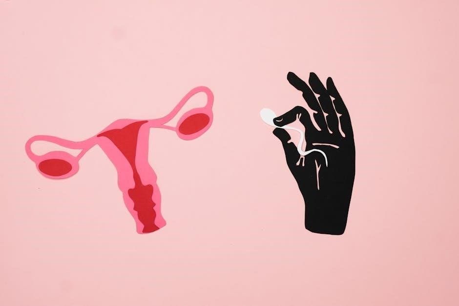
Female Reproductive Anatomy: Visual Guide
Visualizing the female reproductive system starts with external genitalia‚ leading internally to the vagina‚ uterus‚ and fallopian tubes – pathways for egg transport․
The ovaries produce eggs and hormones‚ crucial for reproductive cycles․ Detailed diagrams illustrate the uterus’ layers and the complex structure of the fallopian tubes․
Understanding these components‚ their connections‚ and cyclical changes requires comprehensive visual resources‚ like anatomical atlases and 3D models․
Anatomy and Art: A Visual Synergy
Anatomical knowledge empowers artists to realistically depict the human form‚ mastering both structure and function through detailed‚ knowledgeable visual representations․
Using Anatomical Knowledge for Artistic Representation
Artists benefit immensely from a solid grasp of anatomy‚ enabling accurate and expressive depictions of the human body․ Understanding underlying structures – bones‚ muscles – informs form and movement․
This knowledge transcends mere replication; it allows for stylized yet believable representations․
Treating the body as a ‘map’ with organs as ‘countries’ aids visualization‚ fostering a deeper understanding of spatial relationships crucial for artistic success․
Historical Examples of Anatomy in Art
Throughout history‚ artists have meticulously studied anatomy to achieve realism․ Renaissance masters like Leonardo da Vinci famously dissected cadavers‚ resulting in incredibly accurate depictions of musculature and skeletal structure․
These detailed studies‚ akin to mapping a landscape‚ informed their art․
The ‘map-like’ approach to the body – continents as body parts‚ organs as countries – echoes this historical dedication to anatomical precision․

3D Anatomy Software and Resources
3D Human Anatomy Atlas and similar software offer interactive‚ digital visualizations‚ allowing exploration of the body’s ‘map’ in detail․
Exploring Digital Anatomy Atlases
Digital anatomy atlases‚ such as e-Anatomy‚ revolutionize learning by providing interactive 3D models․ These resources allow students to virtually ‘travel’ through the body‚ much like exploring a detailed map․ Users can dissect structures layer by layer‚ revealing the ‘terrain’ of organs and systems․
This immersive experience surpasses traditional static images‚ fostering a deeper understanding of spatial relationships and anatomical complexity․ These atlases are accessible on various devices‚ enhancing learning flexibility․
Benefits of Interactive Visualizations
Interactive visualizations enhance anatomical learning by moving beyond static images‚ offering a dynamic ‘map’ of the human body․ Students can rotate‚ zoom‚ and dissect virtual structures‚ improving spatial comprehension․ This active engagement fosters deeper understanding and retention compared to passive observation․
Such tools cater to diverse learning styles‚ making complex anatomy more accessible and memorable‚ mirroring the benefits of a well-designed geographical atlas․
Visualizing Anatomy for Different Learning Styles
Visual anatomy adapts to individual needs‚ offering varied ‘map’ representations – diagrams‚ 3D models – to accommodate diverse learners and address impairments․
Adapting Visual Aids to Individual Needs
Visual anatomy thrives on personalization; the ‘map’ analogy shifts based on learner preference․ Some benefit from detailed‚ labeled diagrams‚ while others grasp concepts better with simplified‚ color-coded representations․ Interactive 3D models allow exploration at individual paces․
For those with visual impairments‚ tactile models or audio descriptions paired with visual elements become crucial․ Customizing complexity and presentation ensures accessibility and maximizes comprehension for all students․
Addressing Visual Impairments in Anatomy Education
Despite its visual nature‚ anatomy education can be inclusive․ Beyond traditional diagrams‚ tactile models – representing the ‘anatomical map’ in 3D – are invaluable․ Audio descriptions‚ detailing spatial relationships and structures‚ supplement visual learning․
Technology offers screen readers and adaptable interfaces․ Prioritizing multi-sensory approaches ensures equitable access‚ allowing all students to build a robust understanding of the human body’s complex organization․

Future Trends in Visual Anatomy
Virtual and augmented reality will revolutionize anatomical study‚ offering immersive ‘map’ explorations of the human body‚ enhancing spatial understanding․
Virtual Reality and Augmented Reality Applications
VR and AR transform anatomy education‚ moving beyond static images․ Imagine exploring the body as a 3D ‘continent‚’ dissecting virtually‚ and overlaying anatomical structures onto a patient – a living ‘map․’ These technologies offer immersive experiences‚ improving spatial comprehension and procedural skills․ Students can navigate complex systems‚ like the cardiovascular network‚ as if physically present‚ fostering deeper learning and retention․ This interactive approach caters to diverse learning styles‚ making anatomy accessible and engaging․
The Role of Artificial Intelligence in Anatomical Visualization
AI is revolutionizing anatomical visualization‚ enhancing the ‘map’ of the human body․ Algorithms can generate personalized 3D models‚ highlighting specific structures based on individual needs; AI-powered tools assist in identifying anomalies‚ acting as a ‘guide’ through complex anatomy․ Furthermore‚ AI can create dynamic simulations of physiological processes‚ illustrating function alongside form‚ offering an unprecedented level of detail and understanding․